Corneal Foreign Body Removal at Medivet Godstone Referrals
Peggy- 1 year female neutered DSH
Peggy was seen by her local vet for a sore eye. A corneal foreign body with a penetrating thorn was quickly identified, and she was referred to MGR as an emergency. Although the offending thorn was small, it was deeply embedded in the cornea, penetrating the anterior chamber. This can make for a tricky removal. Fortunately, there was no iris nor lens contact. Surgery was performed that afternoon, using our operating microscope, and the thorn was gently removed. The cornea was sutured and leak tested. The cornea healed well, and Peggy resumed her exciting adventures outside!
Clinical presentation:
- Right eye mild blepharospasm
- Small amount of mucoid discharge
- Moderate conjunctivitis
- Thorn fully embedded and penetrating the cornea, the tip of the thorn within the anterior chamber had a small amount of pale fibrin adhered to it
- Relatively miotic pupil
Referring vet had given medications prior to transfer:
- Cefovecin injection
- Meloxicam injection
- Buprenorphine injection
Blood testing was normal
Surgery:
Under a general anaesthetic, the right eye was prepared for surgery, including clipping the lateral canthal area of hair, followed by a sterile skin and ocular surface preparation with a diluted iodine and saline protocol. Importantly, lubrication of the eye was maintained throughout the procedure with sterile saline.
A small corneal incision was made immediately adjacent to the embedded thorn, deep enough to enable manipulation of the thorn. Using two needles, positioned perpendicular to the thorn, the end was gently grasped and coaxed back out of the cornea.
A mattress corneal suture (vicryl 8-0) was placed around the thorn site. Using a flood of fluorescein, a siedel test for leaking was performed. A slow leak was found, so an additional simple interrupted corneal suture was place. The leak test was repeated to confirm that the cornea was sealed.
Post op:
- Meloxicam oral cat solution once daily
- Chloramphenicol eye drops 4 times daily
- Ketorolac trometamol eye drops 3 times daily
- Remend corneal gel 3 times daily
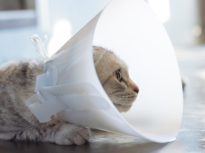
*Buster collar and indoor only for 1 week
Re-checks done at day 4 and 11:
Peggy had a smooth and comfortable recovery. There was a small amount of fluorescein stain uptake at the ends of the corneal suture knots at each visit, but this was very superficial. The rheumocam and acular were stopped at day 11. The owner could not return for a final check, but reported that all was well. The chloramphenicol and remend was stopped a week later.
Corneal foreign bodies are not uncommon with our inquisitive pets. However, they are very serious and need to be addressed urgently. Where there is a shallow corneal penetration of a thorn or piece of metal, careful surgical removal and supportive medications may be adequate. In deep corneal penetrating or perforating foreign bodies, surgical removal is complex and requires magnification, advanced surgical kit and specialised skills. Referral to an ophthalmologist is strongly recommended. If the foreign body has penetrated deep into the anterior chamber, there is a risk of iris or lens penetration. This carries further potential risks of significant uveitis and future cataract development.
For Peggy, a small scar is expected where the cornea was penetrated by the foriegn body. This is unlikely to have any significant effect to her vision and there are no other long-term concerns for Peggy’s eye health.
-
Previous
-
Next


